Structural Methods4 min read
Reading Peptide NMR Structures: Assignments, Restraints, and Ensembles
A peptide NMR structure is an interpretation built from experimental information and a structural model. The familiar image of overlapping conformers is only one part of the record. Understanding it requires the assignments, restraints, validation information, and solution conditions that produced the model.
A peptide NMR structure is an interpretation built from experimental information and a structural model. The familiar image of overlapping conformers is only one part of the record. Understanding it requires the assignments, restraints, validation information, and solution conditions that produced the model.
For researchers comparing peptide structures, the central question is not which image looks most compact. It is how the available measurements support the proposed conformation and what remains uncertain.
Separate assignments from structural conclusions
Chemical-shift assignments connect observed resonances with atoms in the molecule. Additional experimental observations can provide information used as structural restraints. These are related stages, but an assignment list is not itself a complete three-dimensional structure.
The Biological Magnetic Resonance Bank standards resources explain conventions for chemical shifts and related NMR data. Preserving these conventions matters when comparing records generated by different laboratories.
Check that the sequence, residue numbering, and modifications in the assignment record match those in the structural model. A numbering offset can make an apparently local disagreement difficult to interpret.
Read the ensemble with appropriate caution
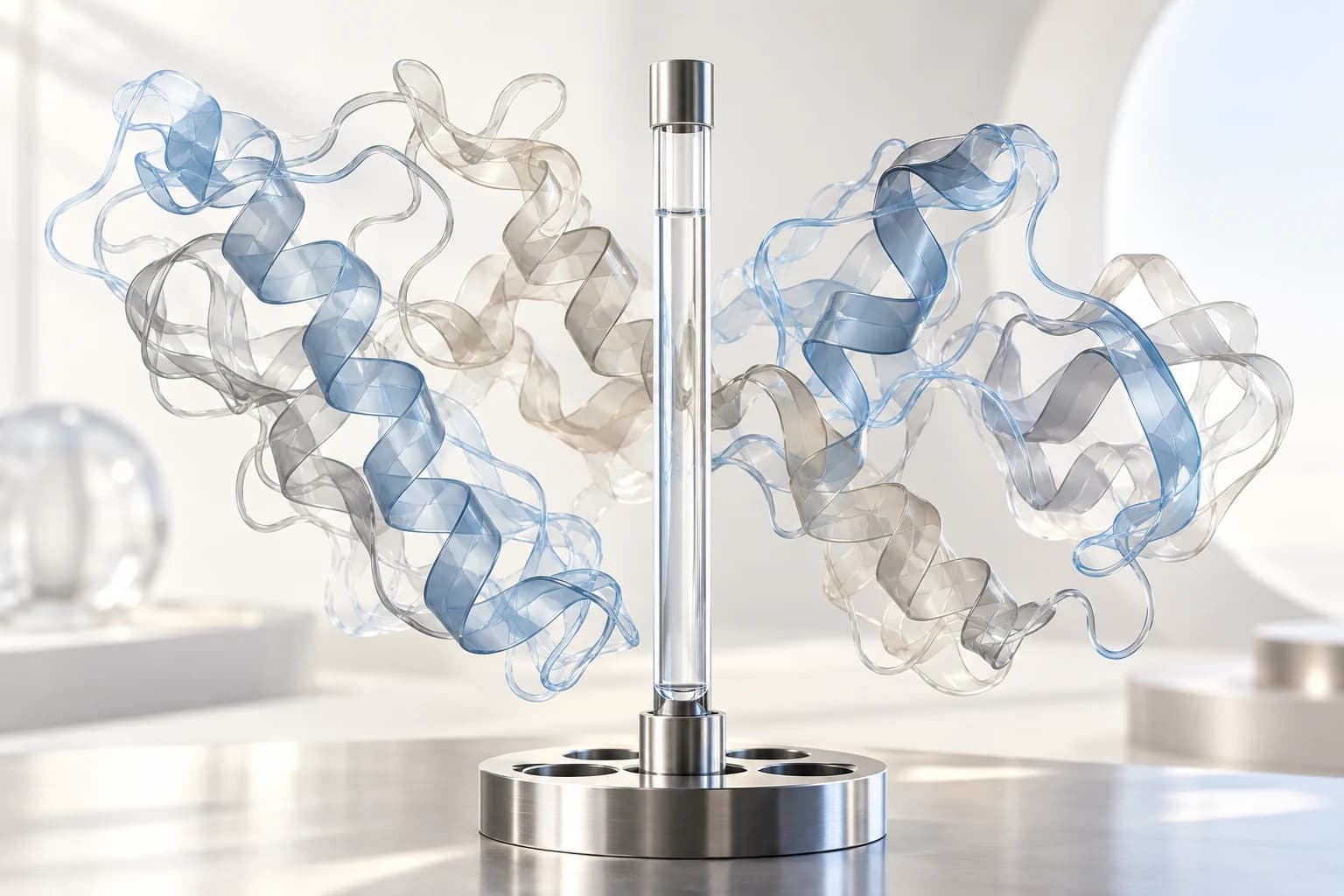
NMR results may be represented by an ensemble of models consistent with the available restraints and calculation process. RCSB PDB's explanation of structure determination places NMR models in the context of experimental structural biology.
Spread among conformers should not automatically be converted into a measured population distribution or a movie of molecular motion. It can reflect limitations of the restraints, the modeling approach, and aspects of molecular flexibility.
Ask which region was used for structural alignment. A well-defined core and a poorly constrained terminus can look very different depending on how the ensemble is displayed.
Inspect the restraint support
Identify which regions have direct experimental support and which depend more heavily on modeling assumptions. The quality question is local as well as global: a favorable overall summary can coexist with uncertainty at the site most relevant to the research hypothesis.
In a hypothetical comparison, two models might agree over most of the backbone but differ at a proposed interaction surface. Before attributing that difference to the peptide, check whether the local observations distinguish the alternatives.
Keep violations, ambiguous assignments, and unrestrained regions visible in the review. Removing inconvenient models from a figure without documenting the selection changes how the evidence appears.
Preserve the solution context
Record the solvent system, pH information, temperature, concentration, and molecular form associated with the experiment. A structure observed under one set of conditions should not silently become the assumed conformation in a different assay environment.
When comparing studies, make a small condition table before comparing coordinates. This helps distinguish a genuine scientific difference from a comparison between different materials or experimental settings.
The site's sequence and modification resource supports the material side of that record. It does not substitute for the NMR acquisition and modeling details.
Connect the model to the next experiment
An NMR structure can guide questions about conformation or interaction, but those questions still require suitable experimental tests. A visually plausible contact in a model does not establish receptor activation, binding strength, or a biological outcome.
For a practical handoff, provide the structure accession where available, assignment source, restraint reference, modeling software information, and the precise question the model is being used to address. Keep the original model files alongside any figures prepared for discussion.
For product-related records, contact Peptide Wholesale Supply about the specific material. Availability of a published structure does not establish that a supplied sample has been characterized by NMR or that the publication studied that sample.
Related reading
Continue with Circular Dichroism for Peptide Secondary Structure: Reading the Spectrum; HDX-MS in Peptide Interaction Research: Reading Protection Changes; Peptide Structures in the PDB: Checking Coordinates and Biological Context.
For in vitro laboratory research use only. Not for human or animal use or consumption. Not for diagnostic or therapeutic use.